A blow to a bent knee can damage the PCL alone or form part of a more serious multiligament injury. You will learn how to recognise urgent warning signs, understand the examination and imaging process, compare rehabilitation with surgery, and prepare for a useful local consultation.
Key takeaways
- Seek emergency care after a knee dislocation, deformity, numbness or a cold foot.
- A dashboard impact can tear the PCL without causing immediate severe swelling.
- MRI shows PCL damage; X-rays check fractures and bone alignment.
- Rehabilitation treats many isolated PCL injuries; combined tears may require surgery.
How PCL injuries happen and when the knee needs emergency care
A PCL injury commonly follows a dashboard-type blow to the front of a bent knee during a vehicle accident, a fall directly onto a bent knee, forceful hyperflexion, or a sports collision. A knee dislocation can also stretch or tear the PCL.
High-energy trauma raises concern for combined damage to the ACL, MCL, posterolateral corner, meniscus, cartilage, bone, blood vessels or nerves.
Arrange a routine orthopaedic appointment for pain, swelling or instability when the knee remains aligned and you can walk safely. Seek immediate assessment for:
- A visibly deformed knee or suspected knee dislocation.
- Inability to bear weight.
- A cold, pale or numb foot.
- An absent foot pulse.
A dislocated knee can reduce by itself before you reach a hospital, making the joint look normal. The popliteal artery and nearby nerves can still be injured, so a reduced knee does not make the event safe to ignore.
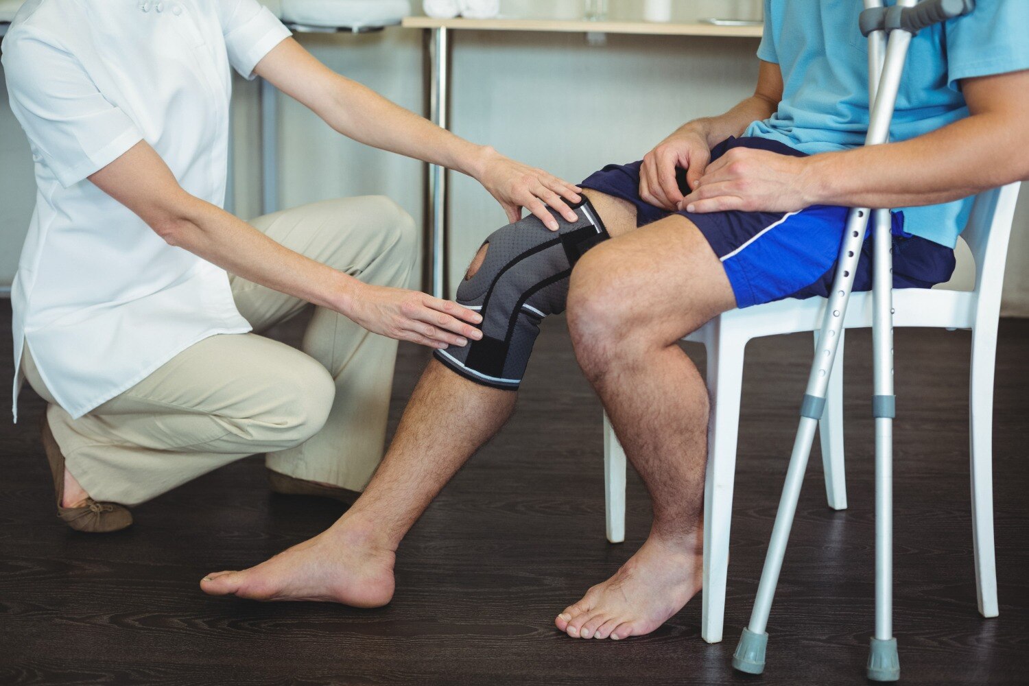
Before consultation, stop pivoting, running and contact sports. Apply ice wrapped in cloth for 15 to 20 minutes, elevate the leg, and use crutches if weight-bearing hurts. Avoid forceful stretching and massage; both can worsen an unstable injury.
If you are searching for PCL treatment in Vile Parle or PCL treatment Mumbai, emergency signs take priority over arranging a routine clinic visit.
How PCL symptoms differ from an ACL tear, meniscus injury or fracture
Posterior knee pain, swelling, painful walking, instability on stairs, difficulty decelerating, or a knee that gives way can indicate a PCL injury, even when early symptoms feel mild. Because the PCL limits backward movement of the tibia, its failure creates a backward sag or shift rather than the forward-slipping pattern associated with an ACL tear.
| Condition | Typical clue | Instability or warning pattern |
|---|---|---|
| PCL injury | Posterior pain, swelling and difficulty slowing down | Tibia shifts backward; stairs and walking feel insecure |
| ACL tear | Pop, rapid swelling and trouble pivoting | Knee feels unstable with cutting or sudden direction changes |
| Meniscus injury | Joint-line pain, catching or locking | Motion becomes blocked or painful at a specific angle |
| Patellar instability | Kneecap feels as if it moves sideways | Shifting occurs at the front of the knee |
| Fracture | Severe pain, marked swelling or tenderness | Deformity or inability to bear weight needs urgent assessment |
Grade I means a PCL sprain, Grade II means a partial tear, and Grade III means a complete tear with significant laxity. A high-energy crash or collision raises concern for combined damage to the ACL, MCL, posterolateral corner, meniscus, cartilage or bone, not an isolated PCL tear.
Someone searching “PCL injury doctor Vile Parle” should ask whether the examination identifies combined injury and whether symptoms fit posterior laxity rather than relying on pain severity alone. A mild start does not rule out instability that progressively changes knee loading.
What the orthopaedic consultation and imaging can reveal
Diagnosis does not rest on MRI alone. During a PCL consultation near me, the clinician compares both knees, reviews how the injury occurred, and checks whether instability affects walking, stairs or deceleration.
The physical examination estimates posterior laxity and can reveal injuries that a scan might not explain by itself:
- Posterior drawer test: the examiner pushes the tibia backward with the knee bent and compares its movement with the uninjured side.
- Posterior sag sign: the tibia sits backward under gravity, suggesting loss of PCL restraint.
- Quadriceps active test: tightening the quadriceps pulls the tibia forward from a sagged position, supporting PCL deficiency.
- Varus and valgus testing: inward or outward stress checks the lateral and medial collateral ligaments.
- Rotational stability testing: abnormal external rotation can point to posterolateral-corner injury.
X-rays do not directly show a midsubstance PCL tear, but they can expose an avulsion fracture, another fracture, dislocation, alignment problem or degenerative change. An avulsion differs from a midsubstance tear because the ligament remains attached to a fragment of bone that has pulled away.
MRI assesses PCL continuity or oedema, posterior translation of the tibia, menisci, cartilage, bone bruising and damage to other ligaments. CT is mainly reserved for complex fractures or dislocations when detailed bone anatomy will change management. A posterior cruciate ligament specialist combines these findings rather than treating an MRI image in isolation.
When rehabilitation is appropriate and when surgery enters the discussion
An isolated, stable Grade I sprain or partial Grade II PCL injury can often begin without surgery. The usual route combines a brace, activity modification, and pain and swelling control before progressive rehabilitation restores motion and function.
| Option | Main approach | When it applies |
|---|---|---|
| Rehabilitation | Quadriceps strength, knee control, balance, motion, and gradual work- or sport-specific tasks | Stable isolated Grade I or partial Grade II injury |
| Reconstruction | Graft-based restoration of the PCL, with treatment of other damaged structures | Persistent instability after structured rehabilitation, severe tear, or combined ligament injury |
| Fixation | Reattachment of a bone fragment to restore the ligament’s origin or insertion | Displaced PCL avulsion or associated fracture |
Early rehabilitation limits posterior tibial sag and excessive hamstring-driven posterior translation. Advancing too quickly can preserve instability, so demanding work, pivoting, and sport wait until strength and control return.
A displaced avulsion is not the same as a midsubstance tear: the PCL remains attached to a bone fragment that has pulled away. Fragment displacement, knee stability, and fixation feasibility guide treatment. For reconstruction, graft choice, tunnel placement, and single- versus double-bundle technique are planning decisions, not automatic proof that one method suits everyone.
A posterior cruciate ligament specialist can explain these choices during PCL treatment Mumbai consultations. Return to demanding activity takes months, not days.
How to choose a PCL consultation in Vile Parle and prepare for it
Choose a PCL consultation in Vile Parle that explains the injury, not just the MRI report. The assessment should establish whether it is acute or chronic, isolated or combined, and associated with an avulsion, fracture, meniscal injury or cartilage damage.
| Question | Useful answer | Warning sign |
|---|---|---|
| Acute or chronic? | Dates the injury and identifies ongoing laxity | No timeline is recorded |
| Isolated or combined? | Checks the ACL, MCL, posterolateral corner, meniscus and cartilage | Only the PCL appears on the checklist |
| Imaging plan | Uses X-rays for fracture or avulsion and MRI for soft-tissue injury | MRI is treated as the entire diagnosis |
| Treatment route | Links stability and symptoms to rehabilitation or surgery | Surgery is recommended without examination findings |
Bring these details to the appointment:
- Injury date, mechanism and whether the knee twisted, struck the dashboard or struck the ground
- Current ability to bear weight, climb stairs and control deceleration
- Previous knee problems, operations and medication list
- Every X-ray or MRI image, plus the written reports
Ask what the posterior drawer grade means, whether the knee is stable enough for rehabilitation, which brace and activity limits apply, and what finding would make surgery necessary.
Dr Dhirav Shah- Orthopaedic Surgeon 8104619499 can assess these findings and discuss whether non-operative care, further imaging or surgical referral fits your case when comparing pcl consultation near me, pcl injury doctor vile parle or pcl treatment in vile parle.
Related service
PCL Treatment PCL or posterior cruciate ligament work opposite to the ACL and located at the back of the knee connecting the femur and the tibia View service → |
Frequently asked questions
When does a PCL injury need emergency care?
Seek emergency care after a knee dislocation, visible deformity, loss of sensation, severe swelling, or a cold or pale foot. These signs can indicate blood-vessel or nerve injury.
How does a PCL tear differ from an ACL tear or meniscus injury?
A PCL tear often follows a blow to the front of a bent knee and causes pain, swelling and backward looseness. ACL tears commonly follow twisting, while meniscus injuries cause joint-line pain, catching or locking. X-rays and MRI help separate these injuries from fractures.
What happens during a PCL consultation?
The orthopaedic examination checks swelling, range of motion and backward movement of the shin. X-rays assess fractures and alignment; MRI shows the PCL and associated ACL, meniscus or cartilage damage.
When is PCL surgery considered?
Rehabilitation is appropriate for many isolated, stable PCL injuries. Surgery enters the discussion when the ligament is completely torn with persistent instability, when multiple ligaments are injured, or when symptoms continue despite structured rehabilitation.
How should I prepare for a PCL consultation in Vile Parle?
Bring injury details, prior scan reports, medication information and a list of symptoms. Note when swelling began, which movements cause giving way, and whether you used a brace or crutches.
Keywords